If you're interested in using any of these photographs or this content in any way, please contact me. Send an e-mail to naturalhistoryphotos(at)gmail.com. Thanks!
Showing posts with label veliger. Show all posts

Saturday, February 6, 2016

Little swimmers

Remember the Purple Sea Snail (Janthina umbilicata) egg capsules?


When I took this picture, the embryos were still developing inside the capsules.  A few days later, some of the larvae emerged from the capsules as active swimming veligers.

These Janthina larvae are extremely small, and are good swimmers.  Here are two still shots (the best I could manage), but at the end of this post there's a short video clip that's much better.

View from the side:


View from above:


Mostly what you're seeing is the reddish shell and the ciliated velar lobes.  In the video, watch for the long cilia beating along the edges of the velar lobes the motion is useful for both swimming and feeding.

Note that this video was made under a microscope — it's magnified 200x!

By the way, from what we can gather, not many people have filmed Janthina larvae, so although this is a short video, you're probably one of the few people in the world to watch a swimming Purple Sea Snail veliger! 

[If you're receiving this via e-mail, click on the title of the video below to watch the clip.] 


Tuesday, July 21, 2015

Working on wings to fly

A few days ago, Kate and her crew noticed an intriguing animal in their plankton sample.


We could tell that it was snail veliger (larva), but we weren't certain about the species.

In between broad wings, it had a very pretty shell:


A tapering foot was visible below:


The veliger had large velar lobes and the lobes were edged with bright orange polka dots.  With active bundles of cilia, it was using the velar lobes to swim.  (This is best viewed in the video at the end of this post.)


As mentioned, we weren't familiar with this snail, so we started sending pictures around for help.  No one responded right away, so Eric decided to keep it in the lab for a few days. 

A couple of days later, the veliger had undergone metamorphosis into a juvenile snail!  This made it much easier to identify.


Note that the ciliated velar lobes are gone; the orange and gold speckles have expanded; and there are two siphons the anterior one (to the left) is formed by a flap near the front end, and the back one (to the right) is formed by the folding of the wings.  (In this case, the wings are extensions of the foot.)  Water is pulled into the front siphon, passes over the gill, and then is expelled out the back siphon.

As the snail glided along and explored its environment, we occasionally caught a glimpse of its two dark eyes:


Pacific Stomach Wings (Gastropteron pacificum) are capable of crawling and swimming.

Eric was fortunate to have filmed this snail when it was still a larva (swimming with its velar lobes) and after it metamorphosed into a juvenile.

Check out his video below!  (Click on the title of the video to see a high resolution version.)


Many thanks to Kate and her team for sharing this wonderful find from the plankton off Bodega Head!
 
P.S.  If any gastropod enthusiasts out there are wondering Pacific Stomach Wings are cephalaspideans, related to headshield slugs and bubble snails.

Saturday, October 25, 2014

Seeing spots

I looked through the microscope and all I saw was spots.  Bright orange spots!


I've seen lots of snail larvae, formally known as veligers, but I'd never seen one with orange spots on its velum!

Here's a closer view (below).  How many orange spots do you count?


We counted at least 13 spots.  This velum is divided into four large lobes (two on each side).  Three of the four lobes have three spots, while the fourth lobe (in the upper right corner in the photo above) has four spots. 

The next image shows the veliger and spots from a slightly different angle.  Note also the cilia along the edge of the velum.  The cilia beat very quickly to help the veliger swim.


Of course we wanted to know the identity of such an intriguing veliger.  However, we couldn't find a match in the Larval Marine Invertebrates of the Pacific Northwest or by doing quick searches online.  We have been narrowing down a possible identity in a somewhat unusual way.  

When I searched for "veliger with orange spots on velum," Google showed us a small portion of text from a book with a description that seemed to match but for which we couldn't see the species name.  Eric kept working on it and eventually figured out that the text was probably referring to a British snail known as Mangelia nebula.  Then he found a paper that described the eggs and larvae of the family of snails (the Turridae) that included this species.  The veligers of some British turrid snails (e.g., see Mangelia nebula below) look very similar to our mystery animal, with spotted velar lobes:


Modified from Lebour, M.V. 1934. The eggs and larvae of some British Turridae. Journal of the Marine Biological Association of the United Kingdom 19: 541-558 


So although we don't know for sure, our best guess right now is that this wonderful polka-dotted veliger could be some type of turrid snail.  [There are a number of turrid snails in California, so we're contacting experts to find out if this veliger could be a match for one of them.] 

To help with the identification, we also tried to get pictures of the snail's shell: 


Why does this veliger have such bright orange spots?  You might think that the orange would make it more visible to predators.  Could the spots be distracting?  Could they draw attention to the edge of the velum, rather than the shell?  Could they somehow look like surrounding particles in the water and help to camouflage the veliger?  Can you think of other reasons it might be valuable for a larval snail to have orange spots?


See below for a short video clip of this tiny snail veliger swimming:


Thursday, September 25, 2014

Lights, cilia, action!


If you liked learning about the atlantid heteropod last night, perhaps you'll enjoy this, too.  

I didn't mention that the heteropods have a larval stage that looks very different than the adult.  And although I've talked about veligers (the larval stage of most marine snails) before, e.g., see post from 11 September 2014, this veliger is noticeably different.


Check out those lobes!  Many veligers have a velum (a lobed, highly ciliated structure surrounding the head that is used for swimming and feeding), but it's unusual for the velum to have 6 lobes.  When we saw this veliger, we knew it was something different right away, just because of the spectacular multi-lobed velum.

Here's a nice diagram of an atlantid heteropod veliger:


Modified from Lalli, C.M. and R.W. Gilmer.  1989.  Pelagic Snails: The Biology of Holoplanktonic Gastropod Mollusks.  Stanford University Press, Stanford, California.


Note that the shell of the veliger is different than the shell of the adult.  It's much more rounded (see below):


But just like the adult, the larva can withdraw completely (velum and all) inside the shell: 


This veliger was discovered by Emily after a plankton tow off Bodega Head on 9 September 2014.  Eric has kept it alive in the lab since then and we've seen it developing and growing.

Thiriot-Quiévreux (1973) provided a helpful table with three stages of development in an atlantid heteropod.  As development proceeds, the shell, velum, eyes, and foot all become more complex (see below).

 Modified from Thiriot-Quiévreux, C. 1973.  Heteropoda.  Oceanogr. Mar. Biol. Ann. Rev.  11: 237-261.


When we first looked at it, the veliger was in Stage II two of the lobes were much smaller than the others, the eye spots were relatively small and rounded, and the foot wasn't that noticeable.

However, within 9 days, we saw dramatic changes as the veliger transitioned to Stage III.  The smaller lobes grew substantially so that all 6 lobes on the velum are now of equal size.  The eye spots have become more visible and are now teardrop-shaped.  And the foot has grown as it develops into a swimming fin.

Below are two images so you can see the changes in the velum (the growth of the smaller lobes).  And then you'll get to watch a video with the veliger in action!

Velum with two smaller lobes (they look like the thumbs on a pair of mittens):


Velum with all 6 lobes of equal size:


And here's the highlight video — it shows the atlantid heteropod veliger actively swimming.  Watch for all of the things discussed abovethe long cilia beating along the edges of the velum, the rounded shell, and the transition from having 2 smaller lobes to all 6 lobes of equal length.  By the way, this veliger is less than 0.5 mm long, so we're thankful that a microscope video camera can provide such nice views of this tiny whirling wonder!


P.S.  We'd like to thank Jeff Goddard for helping us confirm this as a heteropod veliger.

Thursday, September 11, 2014

Not so yucky after all


Here's another highlight from that abandoned crab trap pulled up from about 3 miles offshore of Bodega Head on 7 September 2014.  There was something slimy hanging from it, and if you know me, you know I couldn't resist looking at it.  As I got closer, I heard a voice behind me say, "Yucky, Jackie, yucky!"  It was said in a joking way...and you know it didn't stop me.

Well, I could tell that it was an egg mass, but I couldn't see very much while on the boat.  And I knew that it was just going to dry up on deck.  So I found a small plastic container in my backpack and brought it back to the lab.

The next day I looked at it under the microscope.  The following series of images will let you see what I saw as I zoomed in to look more closely:


Pretty cool, right?  Those are small packets of tiny embryos arranged like beads in long strings, all embedded in one large gelatinous mass.

On 8 September, the embryos were still fairly undeveloped (photos above), so it was hard to say what they might become. 

But I decided to keep them around for a little while to see if they would develop further and provide hints about their identity.  Just a few days later, on 11 September, they looked very different (next two photos): 


Note that each larva has a red spot and two large transparent lobes.  The lobes are called velar lobes and are lined with very active cilia.  They help the larvae to swim and feed.  At this stage the larvae are called veligers.  And we can tell that they belong to a gastropod, although we're not sure which species.  [See bottom of post for an update about the i.d.!]

 
These tiny gastropod larvae are much better appreciated in video format.  This clip shows them actively swimming, first from a distance and then up close.  Note their small shells, and if you look very carefully, you can occasionally see the long cilia beating along the edges of the velar lobes.


Mystery veligers from Jackie Sones on Vimeo.


ADDENDUM (12 September 2014): Jeff Goddard (master of all things related to marine invertebrates) has kindly solved this mystery.  Here's what he says: 

"Those are the veligers of the side-gilled slug Pleurobranchaea californica. At hatching they have a translucent red mantle organ located near the anus, a shell about 0.215 mm long, and a fairly large, powerful-looking velum. Like other pleurobranchs, but unlike almost all other opisthobranchs, P. californica veligers lack an operculum (the protective plate/trapdoor on the back of the foot). This is because of their unique larval development, during which the mantle quickly overgrows the shell, preventing withdrawal of the body into the shell and rendering an operculum useless.

The slugs are large predators/scavengers found on sand or muddy bottoms and are frequently attracted to the bait in crab pots. They lay delicate, curtain-like egg ribbons containing strings of elongate egg capsules, each containing 20 to 25 embryos."


With a quick search of the Internet, I found one good picture of Pleurobranchaea laying eggs

Wednesday, June 18, 2014

Mysterious green mass

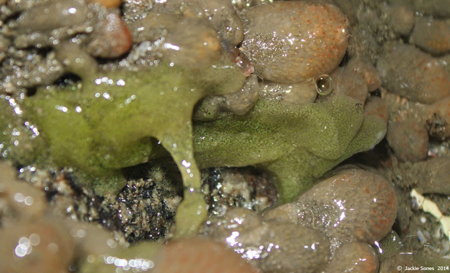
An interesting and mysterious green egg mass in the low intertidal zone along the rocky shore of Bodega Head:


Here's a closeup view (below):


We weren't certain what type of egg mass this was, i.e., what type of animal had produced it.  A worm?  A snail?  So we brought a tiny piece back to the lab for a look under the microscope.  Here's the first view:


Tbe egg mass was packed with developing larvae!  Can you tell what they are?

A couple of the larvae (veligers) had hatched and were crawling on the outside of the egg mass:


Now you can see that this is a tiny gastropod.  The shell is very tall.  Two black eyespots are visible.


The next image shows a nice view from the side:

 
We're not sure which species of gastropod this is, but we'll be asking for ideas.  If you know, let us know!