If you're interested in using any of these photographs in any way, please contact me. Send an e-mail to naturalhistoryphotos(at)gmail.com. Thanks!
Showing posts with label embryo. Show all posts

Wednesday, January 29, 2025

Rose-colored ribbon


A ribbon of rose-colored embryos!  

We were working on a few tasks in the rocky intertidal zone last night (28 January 2025) when Eric came across a Hopkins' Rose Nudibranch (Ceratodoris rosacea) that had recently laid a beautiful string of embryos.

Although we could have missed them in the past, we can't recall finding the embryos of this primarily southern species in Northern California before, so it was an interesting observation for us.

Here's a quick shot of the nudibranch with both its prey (the pink bryozoan, Integripelta bilabiata) and the ribbon of embryos:

 
Thanks for the great photos, Eric!

Monday, July 24, 2023

Turquoise dragon!


Last week, Eric’s summer class collected plankton tows in Bodega Bay and examined them under the microscope.  One of the students (Pachia) spotted this very striking polychaete worm that was turquoise in color and carrying a mass of bright orange eggs!    


The worm was quite small (only ~2.5 mm long).  Those are millimeter marks on the ruler below:  


This worm is known as an epitoke, a type of reproductive individual that occurs in some species of polychaetes.  Epitokes swim up from the bottom to the surface where mass spawning events occur.  In most cases, both male and female epitokes free-spawn their gametes into the water where fertilization occurs.   


However, the species shown here is unusual in that females do not free-spawn, but instead hold onto an egg mass and protect their fertilized embryos for some time while they develop.  Although we aren’t able to identify the species, this worm is likely a member a member of a particular group of polychaetes (Family Syllidae, Subfamily Autolytinae). 


Luckily, Eric was able to capture some video of this beautiful worm, whose appearance reminds us of a tiny turquoise dragon.  [If you can't see the video player in your e-mail, click on the title of the post to go directly to the NHBH website to watch the video.]


With many thanks to Pachia for spotting this wonderful worm, to Bruno and Richard for helping with the identification, and to Eric for putting together the story with photos and video.


Monday, July 4, 2022

The Dove Snail Attacks!

I had a bit of an eyebrow-raising experience today.  I was trying to photograph a common snail, the Carinate Dove Snail (Alia carinata).  This marine snail can be found in a variety of habitats, e.g., rocky shores in tidepools and among surfgrass and seaweeds and in harbors among eelgrass.  It's fairly small (most individuals are probably ~10 mm long), elongate, and usually brownish/black in color.  Here's an example:

 
This species is common enough that you can overlook it.  But I needed a photograph, so I was looking at several of them under a microscope today when I came across one in a puzzling position.  I wasn't sure what was going on at first:
 
 
So that's the snail, with its siphon extended up and to the right and its foot flat against the surfgrass blade (in this photo the foot has blue highlights).  You can see the snail's short tentacles, and you can also see a small dark eye on the right hand tentacle.  But what's below the tentacles?  Here's a side view:
 
 
Ahhh!  The snail had extended its proboscis (a tubular feeding structure) into an egg capsule filled with developing larvae.  It was vacuuming up the veliger larvae!  I hadn't seen this behavior before, so I wasn't sure I was interpreting it correctly at first, but there was no doubt.  The snail reached around inside the egg capsule and cleaned out almost every developing larva.  (The egg capsule/larvae belong to a different snail species, Lacuna marmorata.)
 
Even better, Eric wasn't far away and he came over just in time to capture a video of this fascinating feeding behavior (see below).  
 
I loved learning something new and unusual about a common local snail.  In some ways the name "dove snail" suggests a rather sedate snail, but I'll never look at Alia the same way again!
 
And here's the best part the video!  Watch for the snail approaching the egg capsule, inserting its proboscis inside, then using suction and its tongue-like radula to ingest the tiny larval snails.  You'll see the radula flicking in/out at the tip of the proboscis.  (At this stage, the larval snails have shiny little shells.)


Many thanks to Eric for a memorable video!
 

Sunday, October 4, 2020

Captivating capsules


We came across several squid egg capsules washed up on the beach yesterday (3 October 2020).

These elongate gelatinous capsules contain lots of developing embryos (somewhere between ~50-300, depending on the capsule), each within their own compartment.  It's somewhat similar to a beehive, so if you look carefully at the photo above, you can see that the capsule contains many roughly hexagonal-shaped areas and each one of those has a tiny squid embryo.  (The squid larvae are ~3 mm when they hatch.)

Here's a closer view:

 
Above you can see several embryos, each with two bright red eye spots, a relatively short body section behind the eyes, and a drawn out teardrop-shaped yolk.

When you zoom in even further, you'll notice small chromatophores (pigment spots) on the mantle/main body section!

 
The time it takes for the embryos to develop is dependent on water temperature.  In our area it might take ~45-75 days (faster development in warmer water) before the tiny squid larvae are ready to hatch and swim away from the capsule.

P.S.  If you'd like to see squid embryo chromatophores in action, I posted a short video clip back in 2014 scroll down to the bottom of the post called "Be dazzled!" on 8 October 2014.

Wednesday, August 26, 2020

The things she carries


Check it out!  When sorting through some low intertidal samples looking for ribbon worms, Maddy spotted this tiny polychaete worm.  It was only ~2 mm long, and it was brooding embryos along its back!

For orientation, look for the red eyes at the anterior end.

Before this, we hadn't known that some marine worms use this life history strategy.  (It reminded me of the frogs that brood embryos on their backs.)  The embryos undergo direct development, i.e., they'll continue to develop on their mother's back until they're tiny juvenile worms.

Meet Salvatoria sp.!


Many thanks to Maddy for spotting this fascinating worm, to Eric for taking photos, and to Leslie for help with the identification.

Tuesday, July 28, 2020

All in one basket?


An egg case with developing embryos of a Channeled Basket Snail (Nassarius fossatus).  The capsules are often attached to blades of Eelgrass (Zostera marina).  The embryos hatch in ~20 days.  This capsule was photographed in Bodega Harbor last week.

Tuesday, July 4, 2017

Phoronids for the Fourth

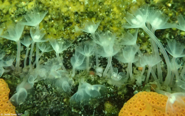
Eric and his summer students spotted these wonderful phoronids, Phoronis vancouverensis, at the Spud Point Marina docks yesterday (3 July 2017):


Remember that phoronids are known for their beautiful lophophores, the U-shaped crown of tentacles shown below:


While looking at these phoronids, we noticed some of the lophophores looked a little differentsome had paired white spots in the center of the lophophore:


Here's a closer look (below).  Note that each of the white spots looks like a little bunch of grapes (clusters of smaller rounded blobs).  Do you have a guess about what they are?


The next image is the closest I could get with my camera at the time:


Each of the white clusters is a group of small developing embryos. Phoronis vancouverensis is a brooder!  The embryos will develop within the lophophore for about 2 weeks.  Then the tiny larvae will swim away, spending time in the plankton before returning to the benthos and undergoing metamorphosis into tiny juvenile phoronids.

I felt fortunate to be photographing phoronids on the Fourth of July!  :)

P.S.  For more information about phoronids, review the post from 27 June 2013.

Wednesday, January 27, 2016

Little red caps

About a week ago, I wrote about Purple Sea Snails (Janthina umbilicata).  We found those snails on 20 January.  A few days later, on 23 January, I found a few more and one of them had eggs!

Janthina lays egg capsules and attaches them to the underside of its float.  In the photo below, it's easy to see the bubbles making up the float on the left, and the egg capsules (filled with embryos!) on the right:


A close-up reveals the small white embryos inside the clear capsules.  One book estimated that Janthina umbilicata has about 80 embryos per capsule.  What do you think?


We kept this snail in the lab, and here's what the embryos looked like today (4 days later):


They've grown quickly!  Their cap-shaped shells are an intriguing shade of brownish-red:


The embryos are still very small (the egg capsules themselves are only ~2 mm long).  This is the best view I could get:


Eventually, the larvae will emerge from the capsules as free-swimming veligers.  But I haven't been able to find information on development time within the egg capsules, or the duration of the larval stage once free of the capsule.  For example, how long does it take until the swimming larvae metamorphose into floating snails and start living at the surface of the ocean?  When do they create their first float?  (How many bubbles are in their first float?  When do the shells become purple?  Does the color change happen when they start eating Velella?)

Thursday, November 12, 2015

The masses

Back in September, while doing a shorebird survey in Bodega Harbor, a couple of different egg masses on the tidal flats caught my eye.

They attracted my attention not only because they were so noticeable, but also because I have been doing shorebird surveys in the same area for over 10 years now and I didn't recall seeing these egg masses before.

One was bright yellow and wrapped around algae or seagrass:


The other was bright white, shaped like a corkscrew, and embedded in the sand:


I finally tracked down the snail making the yellow egg masses.  It was a bubble snail, Haminoea vesicula.  Here are two images to help you visualize how the yellow egg masses consist of rows of developing embryos:


I showed examples of adult bubble snails on 4 June 2015, so check out those pictures here.


I kept meaning to return to find out which species had produced the white corkscrew egg masses, but hadn't found the time.  Today Jeff helped me out he identified the distinctive egg masses as belonging to Rictaxis punctocaelatus.

Below are two close-ups of these spiraled egg masses: 


I haven't seen adult Rictaxis yet (it's been on my wish list!), but if you'd like to see examples of these wonderful local gastropods, check out pictures here.

Thursday, September 11, 2014

Not so yucky after all


Here's another highlight from that abandoned crab trap pulled up from about 3 miles offshore of Bodega Head on 7 September 2014.  There was something slimy hanging from it, and if you know me, you know I couldn't resist looking at it.  As I got closer, I heard a voice behind me say, "Yucky, Jackie, yucky!"  It was said in a joking way...and you know it didn't stop me.

Well, I could tell that it was an egg mass, but I couldn't see very much while on the boat.  And I knew that it was just going to dry up on deck.  So I found a small plastic container in my backpack and brought it back to the lab.

The next day I looked at it under the microscope.  The following series of images will let you see what I saw as I zoomed in to look more closely:


Pretty cool, right?  Those are small packets of tiny embryos arranged like beads in long strings, all embedded in one large gelatinous mass.

On 8 September, the embryos were still fairly undeveloped (photos above), so it was hard to say what they might become. 

But I decided to keep them around for a little while to see if they would develop further and provide hints about their identity.  Just a few days later, on 11 September, they looked very different (next two photos): 


Note that each larva has a red spot and two large transparent lobes.  The lobes are called velar lobes and are lined with very active cilia.  They help the larvae to swim and feed.  At this stage the larvae are called veligers.  And we can tell that they belong to a gastropod, although we're not sure which species.  [See bottom of post for an update about the i.d.!]

 
These tiny gastropod larvae are much better appreciated in video format.  This clip shows them actively swimming, first from a distance and then up close.  Note their small shells, and if you look very carefully, you can occasionally see the long cilia beating along the edges of the velar lobes.


Mystery veligers from Jackie Sones on Vimeo.


ADDENDUM (12 September 2014): Jeff Goddard (master of all things related to marine invertebrates) has kindly solved this mystery.  Here's what he says: 

"Those are the veligers of the side-gilled slug Pleurobranchaea californica. At hatching they have a translucent red mantle organ located near the anus, a shell about 0.215 mm long, and a fairly large, powerful-looking velum. Like other pleurobranchs, but unlike almost all other opisthobranchs, P. californica veligers lack an operculum (the protective plate/trapdoor on the back of the foot). This is because of their unique larval development, during which the mantle quickly overgrows the shell, preventing withdrawal of the body into the shell and rendering an operculum useless.

The slugs are large predators/scavengers found on sand or muddy bottoms and are frequently attracted to the bait in crab pots. They lay delicate, curtain-like egg ribbons containing strings of elongate egg capsules, each containing 20 to 25 embryos."


With a quick search of the Internet, I found one good picture of Pleurobranchaea laying eggs